
|Articles|July 13, 2022
Model digestion: One company describes its new in vitro model for screening how nutrients are absorbed, digested, and made bioaccessible
Author(s)Carolina Galli, Federica Carlomagno
To determine the model’s effectiveness, researchers used the model to examine the chemical effect of digestion in stomach and intestine on a range of nutritional components.
Advertisement
The gastrointestinal tract is a continuous muscular tube which, together with other accessory organs, makes up the digestive system.1 Its main function is to digest food and absorb nutrients and fluids. In addition, it must also protect the body against toxins, pathogens, and other dangerous substances.2 Among all the organs composing the digestive system, the stomach and intestine represent two of the most important organs involved in food breakdown and nutrients absorption.1,3
Understanding the digestive system and its functions inevitably requires some notions about the way in which ingested substances, present in food as macromolecules, are finally made available for absorption in the organism. For this reason, it is important to distinguish between two fundamental concepts: bioaccessibility and bioavailability.
Bioaccessibility is defined as the amount of a compound that is released from the food matrix inside the gastrointestinal tract, which is thus available for intestinal absorption. Bioavailability, on the contrary, indicates the fraction of ingested components that are available to be directly used in the normal physiological activities.4
In pharmacology, the oral route is certainly the preferable method of systemic drug delivery. A drug’s absorption, however, is influenced by several factors, such as composition and volume of gastrointestinal fluids, pH and the relative buffer capacity, digestive enzymes, contraction patterns, and bacterial flora in the gut.5
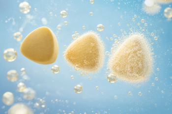
Capsules are one of the most employed solid formats for delivering powders orally. Their content usually consists of active and excipient ingredients, which should not alter the casing; this last one is in fact broken up by digestive fluids in order to let its contents out to be absorbed by the body. Some of the most commonly employed kinds of capsules are DR (delayed release) and HPMC (hydroxyl-propyl-methyl-cellulose) capsules. DR capsules are defined as gastro-resistant, while the HMPC capsules are not.6
Simulating the gastrointestinal tract in vitro by using efficient models could be important in determining the efficacy of a drug or a food supplement in the human body. Several models have been developed at different levels of detail. Some of them are also scientifically recognized. In any case, having a good predictable tool at a lab scale, one that is easy to set up and that can be widely used for any kind of food supplement, should be of great interest to companies as they initially screen new products in development.
Static In Vitro Model Development
This article discusses a recent study that aimed to develop and validate a protocol for assessing the bioaccessibility of food supplements using an in vitro model representative of the human gastrointestinal tract. To determine the model’s effectiveness, researchers used the model to examine the chemical effect of digestion in stomach and intestine on a range of nutritional components. To assess food supplements' bioaccessibility, different products—probiotics, sodium hyaluronate-based, and natural nutraceutical ingredients—were tested in both gastro-resistant and HPMC capsules. Simulated digestive fluids, enzymes (pepsin and pancreatin), and bile salts were employed to reproduce the physiological gastroenteric contents in vitro. (Disclosure: The following model was developed by Roelmi HPC, with an experimental master thesis done with the University of Eastern Piedmont “Amedeo Avogadro”—which are associated with the authors' articles.)
To simulate physiological conditions of the gastrointestinal tract in an in vitro model, literature was deeply screened to determine that it was possible to reproduce the stomach and intestinal environment and their relative internal conditions. As far as gastric and enteric fluids composition were concerned, already-prepared simulated fluids are available on the market, thus providing an easy-to-use mixture which can be previously prepared.7 Enzymes were also added—notably, pepsin and pancreatin concentrations at different values according to retrieved sources.4, 7-15 The same approach was followed for bile salts in intestinal fluid.
Beside fluids composition, other important parameters which needed to be defined included pH, fluid volumes, temperature, stirring velocity, and time (Table 1). In particular, as far as time procedure was concerned, it was important to underline that it is generally standardized to two hours for both gastric and intestinal phases.14-16 However, some authors report 30 minutes as the preferable time for gastric emptying (specifically of capsules), followed then by two hours of intestinal transit. Both the conditions were considered reliable and therefore taken into account in the current study.17
Materials and Methods
Materials
Direct-release (DR) capsules were purchased from Farmalabor S.r.l. (Assago, Italy), while HPMC capsules were supplied by Capsugel S.A.S. (France). The capsules contained different food supplement ingredients, including: 1) probiotics (SynBalance DefensePlus), 2) natural botanical extracts (SelectSieve Rainbow), and 3) sodium hyaluronate (ExceptionHyal Jump). Additionally, a concentrated simulated gastric fluid (SGF) was purchased from Thermo Fisher Scientific Inc. (Rodano, Italy), while concentrated simulated intestinal fluid (SIF) was bought from Reagecon Diagnostics Ltd. (Ireland). Both enzymes (pepsin and pancreatin) were purchased from TCI (Tokyo Chemical Industry Ltd.; Japan). Bile salts were obtained by Liofilchem S.r.l. (Abruzzi, Italy).
Methods
The in vitro digestion procedure was carried out first in a slightly easier way, in order to assess the procedure feasibility. Gastric fluid was complete, while intestinal fluid was at first prepared without pancreatin and bile salts. In a second step, intestinal fluid was prepared also adding enzymes, at different concentrations, and bile salts.
Our procedure required us:
- To prepare the complete simulated gastric fluid solution, diluting the proper amount of concentrated SGF in the correct volume, and adding pepsin in the chosen concentration. This solution needs also to be corrected with a bicarbonate solution to reach pH=2.50±0.05.
- To add the pharmaceutical form to test and set the right timing for the gastric phase, keeping temperature under control.
- In the meantime, to prepare the intestinal fluid solution, diluting the proper amount of concentrated SIF in the correct volume and optionally adding pancreatin and bile salts, in the chosen concentration. Correct pH until a value of 6.50±0.05 was reached.
- Once the gastric phase was over, to pour the already prepared simulated intestinal fluid in SGF.
- To keep the mixture under stirring for two hours, always checking temperature.
- Once the intestinal phase was over, to recover the product and follow different methods for the analysis, according to the kind of ingredient tested.
The methods subsequently employed for ingredients analysis differed depending on the ingredient category. Microscopic evaluation and Gram staining were carried out to assess digestion effects on probiotics. Antioxidant activity was evaluated through DPPH assay for natural ingredients, particularly rich in anthocyanins. Last, a pycnometer and microscopic analysis were performed for sodium hyaluronate–based ingredients to evaluate density.
This detailed procedure has been developed and registered as an internal method.18
Results
The results below refer to the three tested ingredients, with a first visual observation and relative description, especially of fluids aspect at the end of the entire procedure.
Sodium Hyaluronate
With sodium hyaluronate, fluids appeared generally transparent, except when pancreatin and bile salts were added to the intestinal fluid, since they intensified the fluid turbidity. Hyaluronan inside the capsule did not completely dissolve in the fluid but contributed to fluid density increase. In particular, for DR capsules in the procedure without pancreatin and bile salts, the amount of sodium hyaluronate dissolved in the fluid corresponded to 15 mg, while for HPMC capsules it was 120 mg. By contrast, in the complete procedure with all enzymes and bile salts, the amount of the molecule dissolved in the fluid corresponded to 110 mg for DR and 170 mg for HPMC. Lastly, by observing them under the microscope, both before and after the digestion, it was possible to appreciate the difference in hyaluronan chains, which were fragmented into lower molecular weight molecules by digestion simulation (Figure 1).
Probiotics
When probiotics were tested with all enzymes and bile salts, fluids appeared much more turbid, and the powder sediments had a floc-like appearance; conversely, in the procedure with only pepsin, the fluid appeared more transparent and the sediment was quite granular. Moreover, a microscopic analysis was carried out to compare probiotics cells before and after the digestion. After Gram staining, it was possible to appreciate some differences in terms of bacterial cells’ integrity, which was visibly reduced in the procedure involving enzymes and bile salts (Figure 2).
Botanical Extracts
The last product analyzed was a synergy of botanical extracts. The main perceivable result refer to the variation in fluid colour (Figure 3). In fact, this ingredient progressively stained gastric and intestinal fluids from red to violet, also according to the change in pH. Additionally, since this ingredient is rich in anthocyanins, its antioxidant activity through DPPH assay was evaluated. Following the properly adjusted method, absorbance was therefore read for solutions representing pre- and post-digestion, and the obtained values were used to calculate the percentage of DPPH inhibition. The same test was realized also with maqui, a strong antioxidant that was used as a control ingredient. The obtained values for SelectSieve Rainbow were 77.26% before and 44.63% after digestion, while for maqui the percentage of DPPH inhibition was 72.02% before and 31.57% after the procedure.
Discussion
Taking all the above-mentioned results into account, it is possible to affirm that sodium hyaluronate in capsules is progressively cut into lower-molecular-weight fragments (Figure 1), contributing to a significant increase in fluid density. Even though the content did not completely dissolve, the resulting hydrated mass will be probably subjected, in vivo, to muco-adhesion in the intestinal tract, contributing to further degradation and absorption.19
With probiotics, the main outcome refers to the condition in which all enzymes and bile salts were present in the fluid. In fact, a translucent particulate was observed with Gram staining (Figure 2c), which could suggest the presence of broken bacterial cells if observed under direct light (Figure 2d). Effects of bile salts on probiotic survival is known, but partial cell lysis that occurred in the gastrointestinal tract represents the way in which bacterial metabolites are released in the environment, thus contributing to the implementation of their beneficial action in the host.20
It is widely known that polyphenols, like anthocyanins and flavonoids, tend to cause a progressive variation in fluid colour according to pH change21, as it was actually observed for the synergy of botanical extracts (Figure 3). Beside this, the major result obtained for this ingredient was represented by the antioxidant power evaluation through DPPH assay. Although we expected to see a reduction in the scavenging action during the transit in the gastrointestinal tract, it was interesting to find out that the starting antioxidant activity of the synergy was higher than for maqui. This can be explained by the different composition of the two products: differently from maqui, which is mostly made of delphinidins22, the synergy of botanical extract is a mixture of various typologies of polyphenols. They consequently act together to widely implement their antioxidant power.
Conclusion
The aim of this study was to define a protocol to assess the bioaccessibility of different food supplements through the realization of an in vitro model simulating the digestive system at lab scale. With the discrete effect of fluids, enzymes, and bile salts observed on all tested products, it is possible to consider such purpose achieved. Obtained results showed a discrete effect of enzymes and bile salts on all tested products, which was quantified with the proper method of analysis. Such results are encouraging, and they provide room for future research, especially in term of bioavailability evaluation.
Additionally, this study provides the company with a method that can potentially be applied to all of its owned ingredients. Nevertheless, there is still room for future research, especially in the field of bioavailability analysis, which implies a complete investigation of compounds directly released in the bloodstream, ready to be available for normal physiological functions.
About the authors
Carolina Galli represents the Department of Science and Technological Innovation at the University of Eastern Piedmont “Amedeo Avogadro” (Vercelli, Italy). Federica Carlomagno works in the R&D department at
References
- Martini FH et al.
Human Anatomy . Edited by EdiSES s.r.l. (Naples, Italy; 2012) - Fasinu P et al. “
Diverse approaches for the enhancement of oral drug bioavailability .” Biopharmaceutics & Drug Disposition, vol. 32, no. 4 (May 2011): 185-209 - Van de Graaff KM. “
Anatomy and physiology of the gastrointestinal tract .” Pediatric Infectious Disease, vol. 5, suppl. 1 (January-February 1986): S11-S16 - Alegría A et al.
Static Digestion Models: General Introduction, Chapter 1 , in: The Impact of Food Bio-Actives on Gut Health: In Vitro and Ex Vivo Models. Edited by Springer OpenAccess (2015) - Marques MRC et al. “
Simulated biological fluids with possible application in dissolution testing .” Dissolution Technologies, vol. 18, no. 3 (August 2011): 15-28 - Al-Tabakha MM. “
HPMC capsules: Current status and future prospects .” Journal of Pharmacy & Pharmaceutical Sciences, vol. 13, no. 3 (2010): 428-442 - Jantratid E et al. “
Dissolution media simulating conditions in the proximal human gastrointestinal tract: An update .” Pharmaceutical Research, vol. 25, no. 7 (July 2008): 1663-1676 - United States Pharmacopeia and National Formulary (USP 43-NF 38). United States Pharmacopeial Convention; 2019.
- Maeng HJ et al. “
Metabolic stability of D-allulose in biorelevant media and hepatocytes: Comparison with fructose and erythritol .” Foods, vol. 8, no. 10 (October 1, 2019): 448 - Vertzoni M et al. “
Simulation of fasting gastric conditions and its importance for the in vivo dissolution of lipophilic compounds .” European Journal of Pharmaceuticals and Biopharmaceutics, vol. 60, no. 3 (August 2005): 413-417 - Klein S. “
The use of biorelevant dissolution media to forecast the in vivo performance of a drug .” American Association of Pharmaceutical Scientists, vol. 12, no. 3 (September 2010): 397-406 - Jantratid E et al. “
Biorelevant dissolution media simulating the proximal human gastrointestinal tract: An update. ” Dissolution Technologies, vol. 25, no. 7 (August 2009): 21-24 - Charteris WP et al. “
Development and application of an in vitro methodology to determine the transit tolerance of potentially probiotic Lactobacillus and Bifidobacterium species in the upper human gastrointestinal tract .” Journal of Applied Microbiology, vol. 84, no. 5 (May 1998): 759-768 - Hur SJ et al. “
In vitro human digestion models for food applications .” Food Chemistry, vol. 125, no. 1 (March 2011): 1-12 - Wright AJ et al. “
Influence of simulated upper intestinal parameters on the efficiency of beta carotene micellarisation using an in vitro model of digestion .” Food Chemistry, vol. 107, no. 3 (April 2008): 1253–1260 - Fratter A et al. “
Novel micellar system for vitamin D3 oral delivery: Assessment of enteric absorption through a digestion-like in vitro model .” Journal of Drug Delivery Science and Technology, vol. 59 (October 2020) - Grimm M et al. “
Characterization of the gastrointestinal transit and disintegration behavior of floating and sinking acid-resistant capsules using a novel MRI labeling technique .” European Journal of Pharmaceutical Sciences. Published online January 10, 2019. - Internal method ROELMI HPC - DR – PR21.0001 – Digestion model.
- Barbosa de Souza A et al. “
Hyaluronic acid in the intestinal tract: Influence of structure, rheology, and mucoadhesion on the intestinal uptake in rats .” Biomolecules, vol. 10, no. 10 (October 8, 2020): 1422 - Marteau P et al. “
Survival of lactic acid bacteria in a dynamic model of the stomach and small intestine: Validation and the effects of bile .” Journal of Dairy Science, vol. 80, no. 6 (June 1997): 1031-1037 - Aleixandre JL et al. “
The role of UV-visible spectroscopy for phenolic compounds quantification in winemaking .” Published November 2018 in Frontiers and New Trends in the Science of Fermented Food and Beverages. DOI: 10.5772/intechopen.79550 - Escribano-Bailón MT et al. “
Anthocyanins in berries of maqui (Aristotelia chilensis (Mol.) Stuntz) .” Phytochemical Analysis, vol. 17, no. 1 (January-February 2006): 8-14
Advertisement
Related Content
Advertisement


Advertisement
Advertisement
Trending on Nutritional Outlook - Supplement, Food & Beverage Manufacturing Trends
1
The GLP-1 Support Category, Explained
2
Prinova to Highlight Solutions for Solubility and Taste Challenges in Functional Beverages
3
Vitamin K2, MK-4 vs. MK-7: A Formulator's FAQ
4
Sirio Europe Expands FizzyBits Portfolio to Address Consumer Demand for Alternative Formats
5